Basic Life Support (BLS) for Pregnant Women: Maternal CPR, AED, and Team Guidance

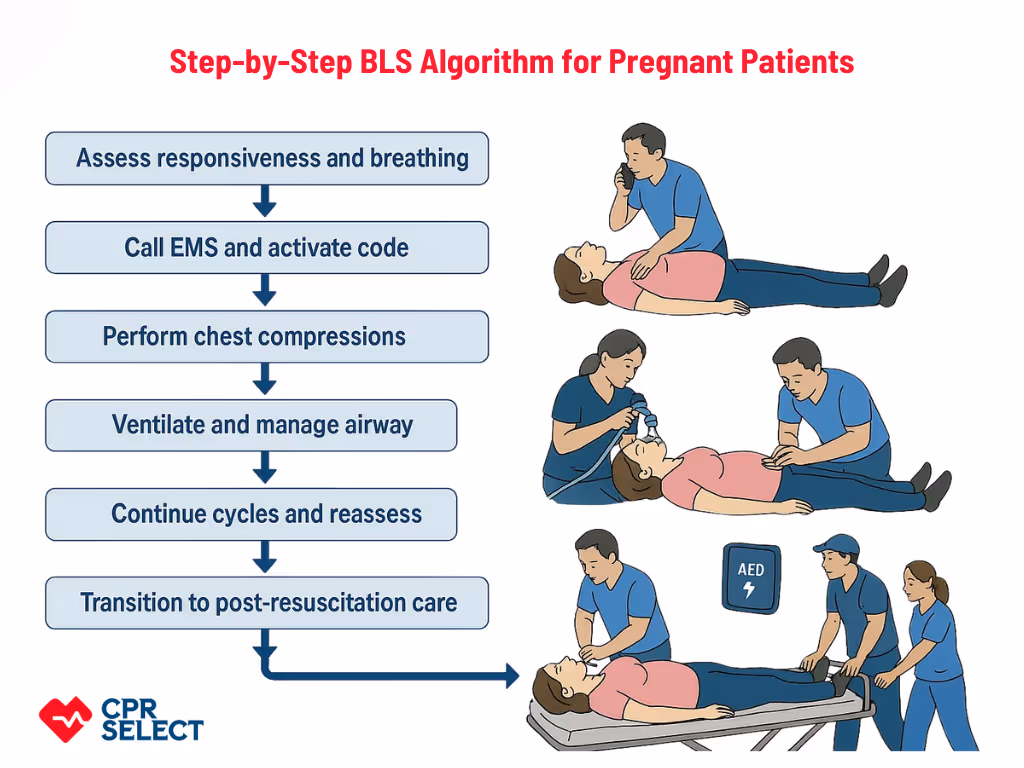
Basic Life Support for pregnant patients adapts standard resuscitation to the physiologic and situational changes of pregnancy, focusing on preserving maternal circulation while considering fetal well being. Pregnancy-specific factors, such as manual left uterine displacement, reduced chest wall compliance, increased blood volume and cardiac output, higher oxygen demand, and the presence of a dependent fetus, affect airway management, ventilation, chest compressions, and resuscitation priorities.

Because maternal circulation is the primary pathway to improving fetal outcomes, BLS for pregnant patients emphasizes restoring effective maternal oxygenation and perfusion first. Responders must understand these core adaptations before learning steps and coordination roles, and specialized BLS training prepares healthcare providers, first responders, trainees, and trained lay rescuers to apply these modifications confidently during emergencies.

Why Basic Life Support Is Critical During Pregnancy?

Why Basic Life Support Is Critical During Pregnancy?

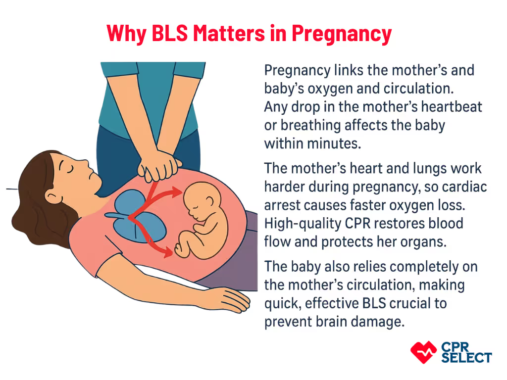
BLS is vital during pregnancy because delays or incorrect actions can quickly harm both the mother and the fetus. Pregnancy creates interdependent maternal–fetal physiology, so any drop in the mother’s circulation or oxygen immediately affects the baby.

  • Maternal risks: Pregnancy changes how the heart and lungs work. Blood volume and cardiac output increase by 30–50%. Oxygen needs rise. Lung capacity drops by about 20%. During cardiac arrest, the mother loses oxygen and blood flow fast. This can cause brain injury, heart damage, and organ failure. High-quality CPR helps restore oxygen and circulation and prevents further injury.
  • Fetal risks: The fetus fully depends on the mother for oxygen and blood flow. When the mother’s heart or breathing stops, the fetus faces low oxygen, poor placental blood flow, and rising acidity. Restoring the mother’s circulation within 4–6 minutes is critical to prevent fetal brain damage. Effective chest compressions and proper ventilation for the mother directly support the baby.

Maternal deterioration threatens both mother and baby within minutes. BLS for pregnant patients must be prompt, skilled, and adapted to pregnancy.

Why pregnancy changes priorities?

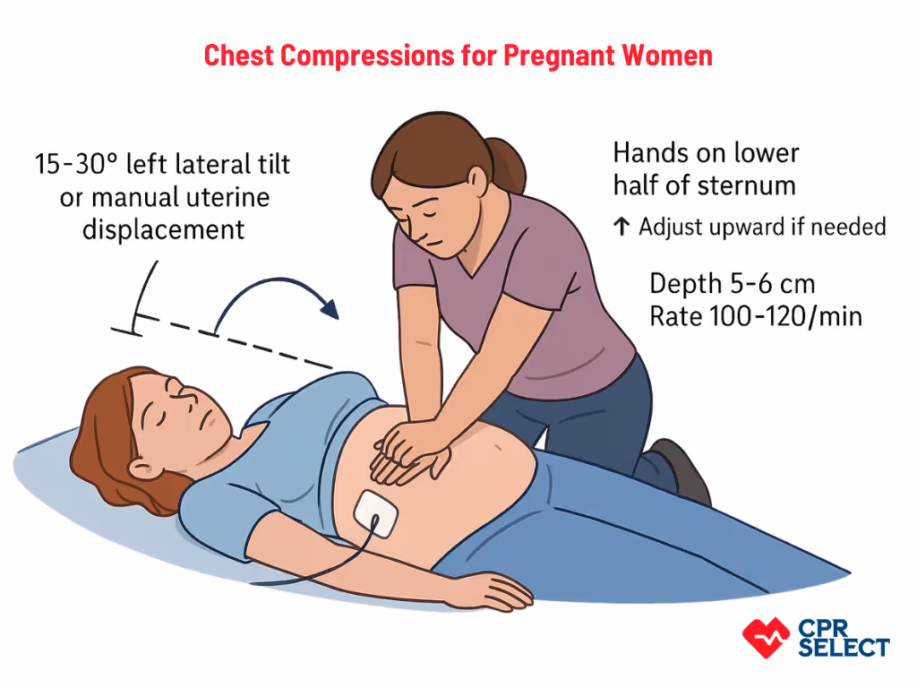
Pregnancy creates a dual-patient emergency. Rescuers must prioritize the mother to protect both lives. The enlarged uterus can compress major blood vessels. For patients beyond 20 weeks, rescuers should use manual left uterine displacement or a 15–30° tilt to improve blood flow while keeping chest compressions effective.

Physiological Changes in Pregnancy Affecting BLS

Pregnancy causes major physiological shifts that affect how Basic Life Support is performed. These changes influence chest mechanics, venous return, airway patency, and oxygen use, so rescuers must adjust standard BLS techniques to maintain maternal and fetal oxygenation. Key physiological changes in pregnancy are as follows:

  • The diaphragm elevates by up to 4 cm.
  • The rib cage widens and shifts forward.
  • Compression hand placement may shift 1–2 vertebral levels higher.
  • Effective compression depth may be harder to achieve.
  • Blood volume increases by 40%.
  • Cardiac output rises by 30–50%.
  • Supine positioning can cause aortocaval compression, reducing venous return and cardiac output.
  • Oxygen consumption increases by 20%.
  • Functional residual capacity decreases by 20%.
  • Oxygen desaturation occurs faster during apnea.
  • Airway tissues become swollen and more prone to bleeding.

These anatomical, cardiovascular, and respiratory adaptations shape how obstetric emergencies present and guide the modified BLS steps.

Common Emergencies in Pregnant Patients

Common Emergencies in Pregnant Patients

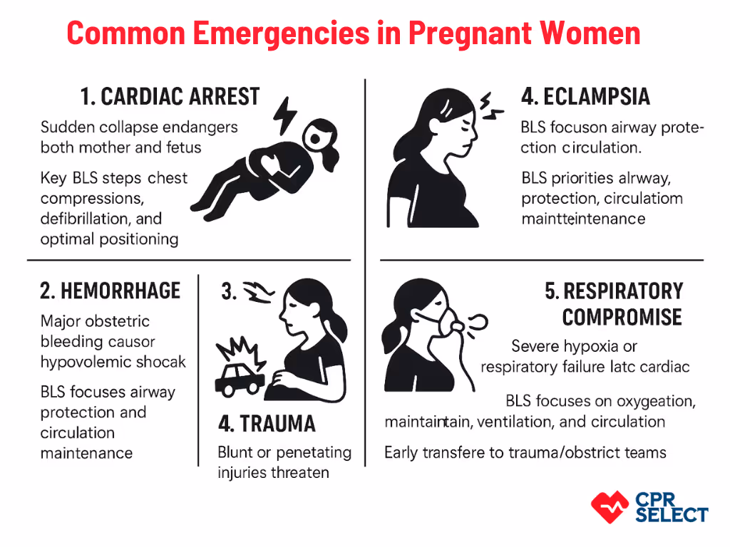
Pregnant patients may face emergencies like maternal cardiac arrest, major obstetric hemorrhage, eclampsia, trauma, and respiratory failure. Each situation presents unique challenges for BLS due to dual-patient considerations, altered maternal physiology, and positional constraints. Rapid recognition and tailored resuscitation are essential to protect both mother and fetus.

1. Cardiac Arrest

Sudden collapse of maternal circulation endangers both mother and fetus. Pregnancy creates a dual-patient scenario and challenges like aortocaval compression, which can reduce venous return by 20–30%. Key BLS steps include high-quality chest compressions (100–120/min, ≥5 cm depth), timely defibrillation, and optimal maternal positioning. If ROSC (return of spontaneous circulation) does not occur within 4 minutes in a viable pregnancy (>20 weeks), perimortem cesarean delivery should be considered within 5 minutes.

2. Hemorrhage

Major obstetric bleeding can cause rapid hypovolemic shock. Immediate priorities include controlling external bleeding, uterine massage if postpartum atony is suspected, preserving circulation and brain perfusion, and activating massive transfusion protocols, while continuing BLS support until definitive care is ready.

3. Eclampsia

Seizures from hypertensive pregnancy disorders can cause sudden loss of consciousness and respiratory compromise. BLS focuses on airway protection, aspiration prevention, left lateral positioning, maintaining circulation, and rapid escalation to critical care. Concurrent management includes magnesium sulfate for seizures and antihypertensives if blood pressure is severely elevated.

4. Trauma

Blunt or penetrating injuries threaten both mother and fetus. Pregnancy-specific considerations include diaphragm displacement, altered anatomy, and potential cervical spine injury. BLS priorities are airway, breathing, and circulation stabilization, spinal precautions, left uterine displacement, and rapid transfer to trauma/obstetric teams.

5. Respiratory Compromise

Severe hypoxia or respiratory failure can lead to cardiac arrest. Pregnancy increases oxygen demand and reduces lung capacity, causing faster desaturation. BLS aims to restore oxygenation, support ventilation, and maintain circulation. Early advanced airway placement and high-flow oxygen are often needed.

Rapid recognition and adapted BLS are critical for all these emergencies. Each condition alters usual resuscitation priorities, and adjustments to chest compressions and positioning are discussed in the next section.

Adjustments to Chest Compressions for Pregnant Patients

Pregnancy alters maternal anatomy and circulation, making chest compressions more challenging. The gravid uterus compresses the inferior vena cava, and thoracic changes affect how compression force is transmitted. These adjustments are essential to maintain maternal cardiac output and fetal perfusion during Basic Life Support (BLS).

Adjustments to Chest Compressions for Pregnant Patients

Left Lateral Tilt

  • Shifts the uterus off the inferior vena cava and aorta.
  • Reduces venous compression and improves cardiac preload and output.
  • Recommended tilt: 15–30° if manual uterine displacement is not possible.
  • Supports both maternal circulation and fetal oxygenation during BLS.

Hand Placement

  • Start on the lower half of the sternum.
  • Adjust slightly upward only if compressions do not produce adequate perfusion or end-tidal CO₂.
  • Consider diaphragm elevation and breast tissue when positioning hands.

Compression Depth

  • Target 5–6 cm, consistent with adult BLS guidelines.
  • Ensure effective systolic pressure while respecting maternal anatomy.
  • Monitor physiologic feedback such as end-tidal CO₂ or return of spontaneous circulation (ROSC).

Compression Rate

  • Maintain 100–120 compressions per minute.
  • Avoid interruptions during airway management or uterine displacement.
  • Coordinate compressions with ventilations and team actions.

This approach ensures high-quality compressions while addressing pregnancy-specific challenges. Coordinating tilt, hand placement, compression depth, and rate is critical to optimize maternal and fetal outcomes.

Airway Management Considerations for Pregnant Patients

Airway management in pregnancy requires awareness of physiological changes that increase airway risk. Rescuers must adapt BLS techniques to reduce aspiration, optimize positioning, and adjust ventilation to maintain maternal oxygenation and fetal perfusion.

  • Pregnancy increases aspiration risk due to higher intraabdominal pressure, delayed gastric emptying, elevated diaphragm, and reduced lower esophageal sphincter tone.
  • Rescuers should prioritize rapid but cautious airway protection and techniques that limit gastric inflation.
  • Elevate the patient’s torso and perform manual left uterine displacement or a 15–30° left lateral tilt to reduce aortocaval compression and improve venous return.
  • Proper positioning also optimizes airway alignment, facemask seal, and laryngeal exposure.
  • Use a two-person bag-valve-mask (BVM) technique whenever possible.
  • Apply cricoid pressure only if trained and after considering possible interference with ventilation.
  • Ventilate with lower tidal volumes (~10 breaths/min) to minimize gastric insufflation.
  • Maintain continuous chest compressions while managing the airway.
  • Assign team members for uterine displacement, facemask seal, and airway escalation to ensure coordination.
  • Airway management should not delay AED use or interrupt high-quality chest compressions.

Effective airway interventions support both maternal oxygenation and fetal perfusion during BLS.

Using an AED on a Pregnant Patient

Using an AED on a pregnant patient is safe and crucial. If a mother experiences cardiac arrest, timely defibrillation can save her life, and indirectly help the baby. AED use is an essential part of Basic Life Support (BLS) for pregnancy and should never be delayed.

When a pregnant patient is unresponsive and not breathing normally, follow standard BLS steps: start CPR immediately, attach the AED, let it analyze the heart rhythm (pause no longer than 10 seconds), deliver a shock if advised, and resume compressions for two minutes. These steps mirror adult BLS protocols but must account for pregnancy-specific factors. Electrode pad placement may need adjustment. You can use:

  • Anterior–lateral: upper pad below the right clavicle, lower pad along the left midaxillary line.
  • Anterior–posterior: one pad on the left midback (level of the inferior scapula), the other on the left anterior chest.
    Make sure the pads stick fully and avoid fetal monitoring devices or external leads. The goal is to maintain current flow through the heart while accommodating the gravid abdomen.

Maternal survival comes first, so defibrillation should never be delayed out of concern for fetal safety. Standard energy settings are safe, and most guidelines emphasize that maternal survival strongly predicts fetal outcomes. AED use should integrate seamlessly with other BLS steps, including continuous chest compressions, ventilations, and manual left uterine displacement if the patient is 20 weeks of gestation or more.

After maternal circulation is restored, focus shifts to post-resuscitation care, including fetal monitoring with nonstress tests or biophysical profiles to ensure ongoing fetal safety.

Using an AED properly on a pregnant patient is life-saving. By combining timely defibrillation with BLS techniques like chest compressions, airway management, and uterine displacement, rescuers can protect both mother and baby effectively.

Special Considerations for Fetal Monitoring

During maternal basic life support (BLS), fetal monitoring is secondary to maternal resuscitation. Its main purpose is to guide obstetric decisions without delaying chest compressions, airway management, or defibrillation. Fetal assessment becomes most relevant at or beyond 20 weeks of gestation, when the gravid uterus can affect maternal circulation and fetal viability may influence resuscitation priorities.

When to consider fetal assessment?

Monitor the fetus only if maternal hemodynamics are temporarily stable or if maternal return of spontaneous circulation occurs. Many guidelines suggest perimortem cesarean delivery if maternal cardiac arrest persists beyond four minutes and the uterine fundus is above the umbilicus. Fetal monitoring should never delay these critical interventions.

How to perform fetal assessment?

Assign a specific team member, such as an obstetric nurse or emergency physician, to use a handheld Doppler, portable ultrasound, or cardiotocograph. Focused point-of-care ultrasound should only check for fetal cardiac activity, lasting a few seconds. Continuous monitoring is not required and must not interrupt BLS tasks.

Interpreting fetal findings

Sustained fetal heart activity indicates potential benefit from continued maternal resuscitation and preparation for neonatal support. Absence of fetal cardiac activity is a poor prognostic sign but should never divert resources from maternal care.

Team coordination

Clearly assign roles for compressors, airway managers, and fetal monitors. Fetal status updates should be concise, such as “fetal heart present at X bpm” or “no fetal cardiac activity detected.” Any decision to escalate to operative obstetric intervention should come only from the team leader after confirming maternal priorities.

Team Dynamics During Maternal Resuscitation

Effective team dynamics are crucial during maternal resuscitation. Clear role allocation and communication reduce delays, prevent overlap, and allow simultaneous attention to maternal and fetal needs.

  • Team leader: Directs the resuscitation, sets priorities, assigns roles, and maintains situational awareness. Gives clear commands such as “Activate obstetrics now” and ensures smooth handovers.
  • Airway manager: Handles airway interventions (bag-mask ventilation, intubation, or supraglottic devices). Coordinates with compressors and ventilators, requests equipment, and confirms placement with capnography when available.
  • Chest compression providers: Perform high-quality compressions at 100–120/min and proper depth. Announce fatigue at 2 minutes and hand off using closed-loop communication. Synchronize pauses only for rhythm checks.
  • Medication and IV/IO manager: Establishes IV or IO access, prepares and delivers medications, and documents doses and times. Coordinates with the recorder to avoid interruptions during compressions.
  • Fetal/obstetric liaison: Monitors fetal status, communicates gestational age and heart tones, and requests obstetric consultation while keeping maternal care primary.
  • Procedural support: Prepares and hands tools for airway and obstetric procedures. Ensures backup devices are ready and maintains sterility.
  • Recorder/timekeeper: Logs events, medications, rhythm checks, and prompts the leader about time-sensitive actions. Tracks compression intervals and overall timeline.
  • Crowd control and safety officer: Manages scene logistics, keeps the area clear, secures equipment, and calls for additional resources if needed. Ensures room readiness for possible perimortem cesarean delivery.
  • Handoffs and personnel augmentation: Incoming staff give concise handovers stating role, capabilities, and completed tasks. Leader confirms via closed-loop acknowledgment.
  • Debrief and role review: Conduct post-event review to assess communication, role clarity, equipment use, and overall team performance. Assign follow-up actions for improvement.

Clear roles, structured communication, and disciplined handoffs are the backbone of successful maternal BLS. Coordinated teams ensure uninterrupted compressions, effective airway management, timely medication delivery, and obstetric considerations aligned with the BLS algorithm for pregnant patients.

Step-by-Step BLS Algorithm for Pregnant Patients

Step-by-Step BLS Algorithm for Pregnant Patients

The basic life support (BLS) algorithm for pregnant patients guides rescuers through maternal cardiac arrest with pregnancy-specific adaptations. The steps prioritize maternal circulation and airway while integrating fetal considerations and professional coordination.

  1. Assess responsiveness and breathing: Quickly determine unresponsiveness and abnormal or absent breathing to decide whether to start chest compressions and call for help.
  2. Call EMS and activate code: Notify emergency services, state that the patient is pregnant, provide gestational age if known, and request obstetric and neonatal-trained personnel while compressions continue.
  3. Perform chest compressions: Begin high-quality compressions at 100–120/min with 5–6 cm depth. Allow full chest recoil, minimize pauses, rotate providers every 2 minutes, and perform manual left uterine displacement if gestation is ≥20 weeks.
  4. Ventilate and manage airway: Deliver ventilations at a 30:2 ratio with bag-mask ventilation or 10 breaths/min if a supraglottic airway or endotracheal tube is in place. Consider aspiration risk, airway edema, and cricoid pressure only if trained.
  5. Use AED if indicated: Attach AED pads in anterolateral or anteroposterior positions, avoiding interference from breast tissue or the abdomen. Deliver shocks per standard adult energy settings and resume compressions immediately.
  6. Continue cycles and reassess: Repeat compressions and ventilations, performing rhythm checks every 2 minutes or sooner if needed. Maintain manual left uterine displacement for patients ≥20 weeks gestation.
  7. Transition to post-resuscitation care: Handover to advanced life support teams or initiate post-resuscitation maternal care once spontaneous circulation returns, ensuring continuity for maternal and fetal monitoring.

This structured sequence forms the core BLS algorithm for pregnancy, integrating adult BLS practices with pregnancy-specific modifications to optimize maternal and fetal outcomes.

Why CPR Select's BLS Certification Prepares Providers?

CPR Select's BLS Certification equips healthcare providers to manage maternal-patient emergencies by addressing gaps in standard BLS knowledge. It teaches maternal-specific skills, including modified chest compressions and manual left uterine displacement, and emphasizes team coordination with obstetric and neonatal personnel. Delivered entirely online, the program focuses on theoretical understanding, scenario-based guidance, and decision-making strategies to build confidence in maternal resuscitation. While it does not provide hands-on practice, it serves as a foundational tool to prepare responders for maternal emergencies and complements institution-specific, in-person training for advanced obstetric and neonatal care.

Get CPR Certified in Minutes for as low as $19.95

Join thousands of professionals that have been certified online with us
100% Online Certification
Fast & Convenient
Instant Certification Card
Nationally Accepted
Get Started
5 star
4.87/5.00
from 259,205 reviews

CPR
Certification

Tailored for the community and workplace
$19.95
$24.95
Offer Expires:
date
Comprehensive CPR Training Across All Ages
Choking response training
Recovery position technique course

Is an AED safe for a pregnant woman?

Yes. An automated external defibrillator (AED) is safe and effective. Place pads in standard anterior–lateral or alternative anterior–posterior positions if breast or abdominal anatomy obstructs placement. Avoid overlapping fetal monitoring devices and maintain pad adhesion for effective defibrillation.

How does pregnancy affect chest compression technique?

Hand placement should shift slightly higher on the sternum to accommodate the gravid uterus. Maintain compressions at 100–120 per minute, with a depth of 5–6 cm. Use manual left uterine displacement while ensuring uninterrupted compressions.

Can you give rescue breaths in maternal CPR?

Yes. Deliver rescue breaths at a ratio of 30 compressions to 2 breaths if using bag-mask ventilation. Ensure chest rise without over-ventilating. Use an oxygen mask if available and consider early supraglottic airway placement in difficult airway scenarios.

When should fetal monitoring be performed during maternal resuscitation?

Fetal assessment is secondary and only performed if it does not interrupt maternal CPR. Use handheld Doppler or focused ultrasound after ROSC or transient maternal stability. Assessment informs possible perimortem cesarean delivery but should not delay the Chain of Survival steps. Most guidelines consider monitoring from 20 weeks gestation onward.

How should airway management be modified for pregnant patients?

Pregnancy increases risk of failed intubation and aspiration due to airway edema. Protect air passages and consider early airway assistance. Employ cricoid pressure only if trained, and use a two-person BVM technique when feasible.

What are the maternal and fetal risks during cardiac arrest?

Maternal CPR takes priority because maternal survival predicts fetal outcome. Fetal risk is mainly due to oxygen deprivation during prolonged arrest. Aim for ROSC within 5 minutes; consider perimortem cesarean delivery if maternal circulation is not restored after 4 minutes in patients beyond 20 weeks gestation.

Can bystanders provide CPR to a pregnant patient?

Yes. Bystander CPR should follow standard adult CPR principles. Early compressions and AED use improve maternal and fetal outcomes. Inform Emergency Medical Services (EMS) that the patient is pregnant for timely obstetric support.

What First Aid and oxygen considerations apply in pregnancy?

Provide standard First Aid including airway clearance and oxygen supplementation. If an obstructed airway occurs, follow BLS choking protocols, considering altered anatomy. Use supplemental oxygen if available to maintain maternal and fetal oxygenation.

Does CPR Select offer training for these scenarios?

Yes. CPR Select Online CPR Certification and BLS/CPR for Healthcare professionals cover maternal-specific adaptations, role allocation, AED use, and emergency scenario awareness. Note that the program is online only and does not include hands-on skills sessions.

Who benefits from maternal BLS/CPR training?

Emergency physicians, laboratory and obstetric nurses, paramedics, medical undergraduate students, and other healthcare professionals in hospital, clinic, and prehospital settings. Training improves readiness for maternal cardiac arrest, cardiopulmonary arrest, and critical events like pulmonary embolism or blood clot complications.